Tuesday

Brain circuit that regulates thirst identified

The thirst-regulating circuit is located in a region of the brain called the subfornical organ (SFO).
Howard Hughes Medical Institute scientists have identified a circuit in the brains of mice that regulates thirst. When a subset of cells in the circuit is switched on, mice immediately begin drinking water, even if they are fully hydrated. A second set of cells suppresses the urge to drink.

The thirst-regulating circuit is located in a region of the brain called the subfornical organ (SFO). "We view the SFO as a dedicated circuit that has two elements that likely interact with each other to maintain the perfect balance, so you drink when you have to and you don't drink when you don't need to," says Charles Zuker, an HHMI investigator at Columbia University who led the research. By doing so, the circuit ensures animals take in the right amount of fluid to maintain blood pressure, electrolyte balance, and cell volume. This work, led by postdoctoral fellow Yuki Oka was published January 26, 2015, in the journal Nature.

Zuker's lab is primarily interested in the biology of taste. Their studies have identified the receptors for the five basic tastes (sweet, sour, bitter, uammi and salt), and shown that the nervous system devotes multiple pathways to sensing and responding to salt. These circuits ensure that salt is appealing to humans at low concentrations, but not at high concentrations. "This is how the taste system regulates salt intake, which is very important for salt homeostasis in the body," says Oka. "But this is just one side of the coin. Salt intake has to be balanced by water intake."

The scientists knew a different mechanism must be responsible for controlling an animal's water intake. "There are no concentration changes for water -- water is water," Oka says. "But when you're thirsty, water is really attractive." Zuker and Oka set out to determine how the brain regulated the motivation to drink.

They began their search in the brain region known as the SFO, which shows increased activity in dehydrated animals. The SFO is one of the few regions of the brain located outside the blood-brain barrier, meaning it has direct contact with body fluids. "These cells might then have the opportunity to directly sense electrolyte balance in body fluids," Zuker points out.

Past experiments in which researchers had elecrically stimulated various circumventricular organs in the brain of mice, including the SFO, had yielded inconsistent results. Oka wanted to find out if their were specific cells in the SFO that triggered drinking behavior. By analyzing genetic markers, he identified three distinct cell types in the SFO: one set of excitatory cells, one set of inhibitory cells, and a third population of supporting cells known as astrocytes.

"If these neurons really mediated key aspects in driving the motivation to drink, then their activation should trigger active drinking, irrespective of the degree of fluid satiety," Zuker says. "And if you silence these populations, you should suppress the motivation to drink, even if you are extraordinarily thirsty."

To test these predictions, Oka introduced a light-sensitive protein into cells in the SFO, enabling the scientists to selectively activate those cells in the mice. Using blue light from a laser, they then switched on the excitatory cells in the SFO of mice that had already had plenty to drink. The results were dramatic.

"There is an animal that is happily wondering around, with zero interest in drinking. You activate this group of excitatory neurons, and it just beelines to the water spout," Zuker says. "As long as the light is on, that mouse keeps on drinking." The animals were uninterested in other fluids, but would avidly drink water for prolonged periods, consuming as much eight percent of their body weight. For humans, that would amount to about 1.5 gallons of water, Oka points out.

"It's very exciting," Zuker says. "This circuit informs and directs the mouse into a complex program of actions and behaviors: "I'm thirsty. I need to identify a source of water. I have to go where the water is. I have to begin to consume that water and I have to continue until this signal is suppressed."

The next step was testing the effect of the inhibitory neurons in the SFO. When Oka switched those cells on in thirsty animals, the mice reduced their water intake by about 80 percent. Activating the inhibitory cells did not affect the animals' interest in food or salt, indicating that the neurons specifically regulate water consumption.

The two groups of cells appear to work together to respond to changing hydration levels and maintain fluid balance, the scientists say. "You can imagine this must be a very tightly controlled feedback loop," Zuker says. "As fluid is being consumed, the electrolyte balance is changing, and this is being sensed."

Zuker also points out "the behavior of the mouse is independent of learning, experience, or context," indicating that the thirst-regulating circuit is hard-wired into the brain. Mingyu Ye, another postdoctoral fellow in Zuker's lab also participated in the study. Yuki Oka recently acccepted a position as assistant professor at the California Institute of Technology.

Selengkapnya »»  

Got bees? Got vitamin A? Got malaria? Loss of pollinators increases risk of malnutrition, disease

Bees allow farms to thrive. But new research from UVM and Harvard scientists shows how bees and other pollinators may be crucial to human health too. The study in PLOS ONE presents the first-ever empirical test of how declining pollinators may increase risk of nutrient deficiencies -- with worrisome connections to diseases like measles and malaria, so prevalent in the developing world.
A new study shows that more than half the people in some developing countries could become newly at risk for malnutrition if crop-pollinating animals -- like bees -- continue to decline.

Despite popular reports that pollinators are crucial for human nutritional health, no scientific studies have actually tested this claim -- until now. The new research by scientists at the University of Vermont and Harvard University has, for the first time, connected what people actually eat in four developing countries to the pollination requirements of the crops that provide their food and nutrients.

"The take-home is: pollinator declines can really matter to human health, with quite scary numbers for vitamin A deficiencies, for example," says UVM scientist Taylor Ricketts who co-led the new study, "which can lead to blindness and increase death rates for some diseases, including malaria."

It's not just plummeting populations of bees. Scientists around the world have observed a worrisome decline of many pollinator species, threatening the world's food supply. Recent studies have shown that these pollinators are responsible for up to forty percent of the world's supply of nutrients.

The new research takes the next step. It shows that in some populations -- like parts of Mozambique that the team studied, where many children and mothers are barely able to meet their needs for micronutrients, especially vitamin A -- the disappearance of pollinators could push as many as 56 percent of people over the edge into malnutrition.

The study, "Do Pollinators Contribute to Nutritional Health?" was led by Alicia Ellis and Taylor Ricketts at UVM's Gund Institute for Ecological Economics and Samuel Myers at the Harvard School of Public Health. It appears in the Jan. 9 issue of the journal PLOS ONE.

Diet details matter

The "hidden hunger" associated with vitamin and mineral deficiencies is estimated to harm more than 1 in 4 people around the globe, the scientists note, contributing to increased risk of many diseases, reduced IQ and diminished work productivity. "Continued declines of pollinator populations could have drastic consequences for global public health," the team writes.

"This is the first study that quantifies the potential human health impacts of animal pollinator declines," says Myers. Earlier studies have shown links between pollinators and crop yields -- and between crop yields and the availability of food and nutrients. "But to evaluate whether pollinator declines will really affect human nutrition, you need to know what people are eating," Myers explains. So the new study examined the full pathway from pollinators through to detailed survey data about people's daily diets in parts of Zambia, Mozambique, Uganda and Bangladesh.

"How much mango? How much fish?" says Ricketts. "And from that kind of data we can find out if they get enough vitamin A, calcium, folate, iron and zinc." Then the scientists were able to examine the likely impact a future without pollinators would have on these diets.

Epidemiology meets ecology

And for parts of the developing world, that future could well include "an increase in neural tube defects from folate deficiency or an increase in blindness and infectious diseases from vitamin A deficiency," Myers says, "because we have transformed our landscapes in ways that don't support animal pollinators anymore."

"We find really alarming effects in some countries for some nutrients and little to no effect elsewhere," Ricketts says. On the bleak end of the spectrum, the team projected little difference in Bangladesh, since so many people there are already malnourished. And, at the other end of the spectrum, Zambia should be relatively insulated from this risk. That's because -- though the scientists project reductions in the intake of vitamin A with pollinator declines in Zambia -- "there is so much vitamin A in the diet already that it didn't push very many people below the threshold," Myers explains.

This new study fits into an emerging field of research exploring how the very rapid transformation of Earth's natural systems affects human health. The big picture? "Ecosystem damage can damage human health," Ricketts says, "so conservation can be thought of as an investment in public health."

Selengkapnya »»  

How malaria-spreading mosquitoes can tell you're home

Ring Cardé, a distinguished professor of entomology at UC Riverside, is seen here working on an experiment involving a wind tunnel.
Females of the malaria-spreading mosquito tend to obtain their blood meals within human dwellings. Indeed, this mosquito, Anopheles gambiae, spends much of its adult life indoors where it is constantly exposed to human odor -- from used clothing, bedding, etc. -- even when people are absent.

But is human odor enough as a reliable cue for the mosquitoes in finding humans to bite?

Not quite, reports a team of entomologists at the University of California, Riverside in a research paper published online earlier this month in the Journal of Chemical Ecology. The researchers' experiments with female Anopheles gambiae show that the mosquitoes respond very weakly to human skin odor alone. The researchers found that the mosquitoes' landing on a source of skin odor was dramatically increased when carbon dioxide was also present, even at levels that barely exceed its background level. The researchers suggest, too, was that the mosquitoes use a "sit-and-wait" ambush strategy during which they ignore persistent human odor until a living human is present.

"Responding strongly to human skin odor alone once inside a dwelling where human odor is ubiquitous is a highly inefficient means for the mosquito of locating a feeding site," said Ring Cardé, a distinguished professor of entomology, whose lab conducted the research. "We already know that mosquitoes will readily fly upwind towards human skin odor but landing, the final stage of host location, which typically takes place indoors, does not occur unless a fluctuating concentration of carbon dioxide indicates that a human host is present. It may be that upwind flight towards human odor has more to do with locating a human dwelling, which emits human odor even when its occupants are absent, than locating a feeding site per se."

Cardé explained that mosquitoes, once indoors, conserve their energy by ignoring omnipresent human odor in an unoccupied room. Small increases in carbon dioxide indicate to the mosquitoes the probable presence of a human. This then triggers the mosquitoes to land on human skin.

The findings could help in the design on new types of mosquito control. One take-home message from this work is that studies defining which human odors mediate host finding and which compounds are good repellents need to precisely control exposure to above ambient carbon dioxide -- an experimenter entering an assay room quickly elevates the level of carbon dioxide and thereby alters the mosquitoes' behavior.

The research shows that when it comes to feeding on humans indoors, malaria mosquitoes have developed a striking adaptation to how carbon dioxide affects their landing on human targets in response to skin odor.

"It also would be useful next to see if mosquitoes' response to skin odor is similarly affected by carbon dioxide in outdoor situations and how these interactions play out in human dwellings," Cardé said.

Larvae of Anopheles gambiae can breed in diverse habitats. This mosquito has evolved to search in human dwellings for blood meals to carry out egg production. The mosquito enters houses throughout the night, peaking around midnight and continuing at a high rate until the early morning hours. Following a blood meal, the mosquito often remains in dwellings until it is ready to lay eggs. Mosquitoes also seek refuge inside human dwellings during the day, taking shelter from high daytime temperatures outside.

Cardé, who occupies the A. M. Boyce Chair in the Department of Entomology, was joined in the study by Ben Webster (first author of the research paper) and Emerson S. Lacey.

To conduct their experiments, the researchers used Anopheles gambiae originating from mosquitoes collected in Cameroon. They collected skin odor by using pieces of white polyester gauze worn by Webster in a cotton sock for 4-6 hours before the experiments began. The landing behavior of the mosquitoes in the experiments was recorded with a video camera equipped with night vision.

Selengkapnya »»  

On the ups and downs of the seemingly idle brain

This image shows inhibitory cells abound in the barrel cortex of the mouse, where three main types were labeled to fluoresce in different colors: PV (red), SOM (blue), and 5HT3aR, which includes VIP and NPY, (green).
Even in its quietest moments, the brain is never "off." Instead, while under anesthesia, during slow-wave sleep, or even amid calm wakefulness, the brain's cortex maintains a cycle of activity and quiet called "up" and "down" states. A new study by Brown University neuroscientists probed deep into this somewhat mysterious cycle in mice, to learn more about how the mammalian brain accomplishes it.

In addition to an apparent role in maintaining a baseline of brain activity, the up and down cycling serves as a model for other ways in which activity across the cortex is modulated, said Garrett Neske, graduate student and lead author. To study how the brain maintains this cycling, he found, is to learn how the brain walks a healthy line between excitement and inhibition as it strives to be idle but ready, a bit like a car at a stoplight.

"It is very important to regulate that balance of excitation and inhibition," said senior author Barry Connors, professor and chair of neuroscience at Brown. "Too much excitation relative to inhibition you get a seizure, too little you become comatose. So whether you are awake and active and processing information or whether you are in some kind of idling state of the brain, you need to maintain that balance."

The cycling may seem simple, but what Neske and Connors found in their investigation, published in the Journal of Neuroscience, is that it involves a good deal of complexity. They focused on five different types of cells in a particular area of the mouse cortex and found that all five appear to contribute uniquely to the ups and downs.

Cells in a barrel

Specifically the researchers, including Saundra Patrick, neuroscience research associate and second author, looked at the activity of excitatory pyramidal cells and four kinds of inhibitory interneurons (PV, SOM, VIP and NPY) in different layers of the barrel cortex. That part of the cortex is responsible for processing sensations on the face, including the whiskers.

Neske induced up and down cycles in slices of tissue from the barrel cortex and recorded each cell type's electrical properties and behaviors, such as its firing rate and the amounts of excitation and inhibition they received from other neurons.

The picture that emerged is that all types of interneurons were active. This included the most abundant interneuron subtype (the fast-spiking PV cell), and the various more slowly spiking subtypes (SOM, VIP, NPY). In fact, Connors said, the latter cells were active at levels similar to or higher than neighboring excitatory cells, contributing strong inhibition during the up state.

One way such findings are important is in how they complement recent ones by another research group at Yale University. In that study scientists looked at a different part of the cortex called the entorhinal cortex. There they found that only one inhibitory neuron, PV, seemed to be doing anything in the up state to balance out the excitement of the pyramidal neurons. The other inhibitory neurons stayed virtually silent. In his study, Neske replicated those results.

Taken together, the studies indicate that even though up and down cycles occur throughout the cortex, they may be regulated differently in different parts.

"It suggests that inhibition plays different roles in persistent activity in these two regions of cortex and it calls for more comparative work to be done among cortical areas," Neske said. "You can't just use one cortical region as the model for all inhibitory interneuron function."

From observation to manipulation

Since observing the different behaviors of the neuron types, Neske has moved on to manipulating them to see what role each of them plays. Using the technique of optogenetics, in which the firing of different neuron types can be activated or suppressed with pulses of colored light, Neske is experimenting with squelching different interneurons to see how their enforced abstention affects the up and down cycle.

When the work is done, he should emerge with an even clearer idea of the brain's intricate and diligent efforts to remain balanced between excitation and inhibition.

Selengkapnya »»  

Which 'letters' in the human genome are functionally important?

"In model organisms, like yeast or flies, scientists often generate mutations to determine which letters in a DNA sequence are needed for a particular gene to function," explains CSHL Professor Adam Siepel. "We can't do that with humans. But when you think about it, nature has been doing a similar experiment on a very large scale as species evolve. Mutations occur across the genome at random, but important letters are retained by natural selection, while the rest are free to change with no adverse consequence to the organism."
There are 3 billion letters in the human genome, and scientists have endlessly debated how many of them serve a functional purpose. There are those letters that encode genes, our hereditary information, and those that provide instructions about how cells can use the genes. But those sequences are written with a comparative few of the vast number of DNA letters. Scientists have long debated how much of, or even if, the rest of our genome does anything, some going so far as to designate the part not devoted to encoding proteins as "junk DNA."

In work published in Nature Genetics, researchers at Cold Spring Harbor Laboratory (CSHL) have developed a new computational method to identify which letters in the human genome are functionally important. Their computer program, called fitCons, harnesses the power of evolution, comparing changes in DNA letters across not just related species, but also between multiple individuals in a single species. The results provide a surprising picture of just how little of our genome has been "conserved" by nature not only across species over eons of time, but also over the more recent time period during which humans differentiated from one another.

"In model organisms, like yeast or flies, scientists often generate mutations to determine which letters in a DNA sequence are needed for a particular gene to function," explains CSHL Professor Adam Siepel. "We can't do that with humans. But when you think about it, nature has been doing a similar experiment on a very large scale as species evolve. Mutations occur across the genome at random, but important letters are retained by natural selection, while the rest are free to change with no adverse consequence to the organism."

It was this idea that became the basis of their analysis, but it alone wasn't enough. "Massive research consortia, like the ENCODE Project, have provided the scientific community with a trove of information about genomic function over the last few years," says Siepel. "Other groups have sequenced large numbers of humans and nonhuman primates. For the first time, these big data sets give us both a broad and exceptionally detailed picture of both biochemical activity along the genome and how DNA sequences have changed over time."

Siepel's team began by sorting ENCODE consortium data based on combinations of biochemical markers that indicate the type of activity at each position. "We didn't just use sequence patterns. ENCODE provided us with information about where along the full genome DNA is read and how it is modified with biochemical tags," says Brad Gulko, a Ph.D. student in Computer Science at Cornell University and lead author on the new paper. The combinations of these tags revealed several hundred different classes of sites within the genome each having a potentially different role in genomic activity.

The researchers then turned to their previously developed computational method, called INSIGHT, to analyze how much the sequences in these classes had varied over both short and long periods of evolutionary time. "Usually, this, kind of analysis is done comparing different species -- like humans, dogs, and mice -- which means researchers are looking at changes that occurred over relatively long time periods," explains Siepel. But the INSIGHT model considers the changes among dozens of human individuals and close relatives, such as the chimpanzee, which provides a picture of evolution over much shorter time frames.

The scientists found that, at most, only about 7% of the letters in the human genome are functionally important. "We were impressed with how low that number is," says Siepel. "Some analyses of the ENCODE data alone have argued that upwards of 80% of the genome is functional, but our evolutionary analysis suggests that isn't the case." He added, "other researchers have estimated that similarly small fractions of the genome have been conserved over long time evolutionary periods, but our analysis indicates that the much larger ENCODE-based estimates can't be explained by gains of new functional sequences on the human lineage. We think most of the sequences designated as 'biochemically active' by ENCODE are probably not evolutionarily important in humans."

According to Siepel, this analysis will allow researchers to isolate functionally important sequences in diseases much more rapidly. Most genome-wide studies implicate massive regions, containing tens of thousands of letters, associated with disease. "Our analysis helps to pinpoint which letters in these sequences are likely to be functional because they are both biochemically active and have been preserved by evolution." says Siepel. "This provides a powerful resource as scientists work to understand the genetic basis of disease."

Selengkapnya »»  

Sunday

Live broadcast from inside the nerve cell

Protein degradation by the proteasome in neurons. The proteasomes (grey) of the nerve cell (neuron) are equipped with the regulatory particles at their ends. These structures change their shape depending on whether they have bound (red) proteins which have to be degraded (green) or not (blue).
Neurodegenerative diseases like Alzheimer's, Huntington's or Parkinson's are caused by defect and aggregated proteins accumulating in brain nerve cells that are thereby paralyzed or even killed. In healthy cells this process is prevented by an enzyme complex known as the proteasome, which removes and recycles obsolete and defective proteins. Recently, researchers in the team of Wolfgang Baumeister at the Max Planck Institute of Biochemistry in Martinsried near Munich were the first to observe and structurally characterize proteasomes at work inside healthy brain cells. "When we saw the proteasomes on our screen, we were immediately aware of the major importance of the results," remembers Shoh Asano, first author of the study. The study has now been published in the journal Science.

Scientists estimate that our brain consists of about ten to one hundred billions of nerve cells. In order to fulfill their respective tasks as long as possible, these cells have to constantly control their internal proteins with regard to quality and functionality. Otherwise the proteins might clump together and thereby paralyze or even kill the cells. Once the cell recognizes a defect protein, this is marked for degradation and a kind of a molecular shredder, the so-called proteasome, chops it into pieces that are eventually recycled.

For the first time now, researchers have succeeded in visualizing this process in intact nerve cells, which previously could only be investigated in the test tube. Electron cryo-tomography was essential for obtaining the described images. Hereby, cells are cooled down to minus 170°C in a fraction of a second. In a consecutive step, pictures of the interior of the cells are taken from many different angles, which then are merged computationally into a three-dimensional image.

"First time in intact cells"
 
In the current study, the use of specific technical innovations allowed the researchers to achieve a unprecedented imaging quality, enabling them to distinguish single proteasomes within the cell. "For the first time it is possible to qualitatively and quantitatively describe this important enzyme complex in intact cells," Asano classifies the results. In the following experiments, the scientists focused on the activity of the proteasomes. For the interpretation of the single particles it is important to know that there are cap-like structures, the so-called regulatory particles, attached to the ends of proteasomes (see picture). They bind proteins that are designated to be degraded and thereby change their shape. The scientists were able to distinguish these states and consequently could deduce how many of the proteasomes were actively degrading proteins.

New prospects for the future
 
The conclusion of the researchers: in quiescent nerve cells like the ones used in the actual experiments, only a minority of the proteasomes is active. In detail, the results showed that only every fourth proteasome was actively degrading proteins while the rest idled at the same time. In the future, the scientists want to address the structural changes of the proteasomes under cellular stress as it occurs in neurodegenerative diseases. "This study shows the new possibilities to resolve protein complexes in their entirety in the cell and to study their mutual functional dependencies," Wolfgang Baumeister determines the agenda for the future.


Selengkapnya »»  

First major analysis of Human Protein Atlas is published

An image from the Human Protein Atlas.
A research article published in Science presents the first major analysis based on the Human Protein Atlas, including a detailed picture of the proteins that are linked to cancer, the number of proteins present in the bloodstream, and the targets for all approved drugs on the market.

The Human Protein Atlas, a major multinational research project supported by the Knut and Alice Wallenberg Foundation, recently launched (November 6, 2014) an open source tissue-based interactive map of the human protein. Based on 13 million annotated images, the database maps the distribution of proteins in all major tissues and organs in the human body, showing both proteins restricted to certain tissues, such as the brain, heart, or liver, and those present in all. As an open access resource, it is expected to help drive the development of new diagnostics and drugs, but also to provide basic insights in normal human biology.

In the Science article, "Tissue-based Atlas of the Human Proteome," the approximately 20,000 protein coding genes in humans have been analysed and classified using a combination of genomics, transcriptomics, proteomics, and antibody-based profiling, says the article's lead author, Mathias Uhlén, Professor of Microbiology at Stockholm's KTH Royal Institute of Technology and the director of the Human Protein Atlas program.

The analysis shows that almost half of the protein-coding genes are expressed in a ubiquitous manner and thus found in all analysed tissues.

Approximately 15% of the genes show an enriched expression in one or several tissues or organs, including well-known tissue-specific proteins, such as insulin and troponin. The testes, or testicles, have the most tissue-enriched proteins followed by the brain and the liver.

The analysis suggests that approximately 3,000 proteins are secreted from the cells and an additional 5,500 proteins are located to the membrane systems of the cells.

"This is important information for the pharmaceutical industry. We show that 70% of the current targets for approved pharmaceutical drugs are either secreted or membrane-bound proteins," Uhlén says. "Interestingly, 30% of these protein targets are found in all analysed tissues and organs. This could help explain some side effects of drugs and thus might have consequences for future drug development."

The analysis also contains a study of the metabolic reactions occurring in different parts of the human body. The most specialised organ is the liver with a large number of chemical reactions not found in other parts of the human body.


Selengkapnya »»  

Bad reputation of crows demystified

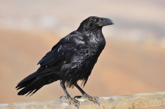
The 326 interactions between corvids and their prey show that they have a much smaller effect on other bird species than was previously thought.
In literature, crows and ravens are a bad omen and are associated with witches. Most people believe they steal, eat other birds' eggs and reduce the populations of other birds. But a new study, which has brought together over 326 interactions between corvids and their prey, demonstrates that their notoriety is not entirely merited.

Corvids -- the bird group that includes crows, ravens and magpies -- are the subject of several population control schemes, in both game and conservation environments. These controls are based on the belief that destroying them is good for other birds. They are also considered to be effective predators capable of reducing the populations of their prey.

However, a study published recently in the journal 'Ibis' analysed the impact of six species of corvid on a total of 67 species of bird susceptible to being their prey, among which are game birds and passerine birds.

The project, which compiled the information of 42 scientific studies and analysed a total of 326 interactions between corvids and their prey, shows that they have a much smaller effect on other bird species than was previously thought.

As Beatriz Arroyo -- author of the study and a researcher at the Institute of Research in Game Resources (IREC), a joint centre of the University of Castilla-La Mancha, the Castilla-La Mancha Community Council and the CSIC (Spanish National Research Council) -- says: "In 81% of cases studied, corvids did not present a discernible impact on their potential prey. Furthermore, in 6% of cases, some apparently beneficial relationships were even observed."

Greater impact on reproduction

To find out what impact corvids have on their prey, the researchers -- in conjunction with the University of Cape Town (South Africa) -- conducted several experiments in which they isolated crows, ravens and magpies, among other predators, to observe how they affected the reproduction and abundance of other birds.

According to the works analysed, when crows were taken away from their habitat, the survival rates of chickens and the number of eggs of other species were higher in most cases. Nevertheless, with respect to abundance, without corvids an increased size of the populations of other birds was observed only in a small number of cases.

According to the study, when crows were removed from the environment, in 46% of cases their prey had greater reproductive success, while their abundance fell in less than 10% of cases.

Additionally, these experimental studies carried out in nine different countries (Canada, France, Norway, Poland, Slovakia, Spain, Sweden, the UK and the USA) revealed that, if corvids are eliminated but other predators are not, the impact on the productivity of their prey would be positive in only 16% of cases; whilst without corvids and other predators, including carnivores, the productivity of other birds improves in 60% of cases.

This suggests that crows, ravens and magpies, amongst others, have a lower impact on prey than other threats. "Compensatory predation can also occur," the researcher explains.

In the study they also compared the effects between different groups of corvids. In these results it is striking that "magpies had much less impact on prey than other species," Arroyo claims.

Comparing crows and magpies, the scientists showed that in 62% of cases crows impacted negatively on the reproduction of their prey, whilst magpies had a negative effect in 12% of cases. "But no differences related to the abundance of prey were noted," the scientist affirms.

For the authors of this piece of research, given the results it is necessary to "be cautious" when drawing conclusions on the impact of magpies or crows on the populations of their prey. "This method of managing populations is frequently ineffective and unnecessary," Arroyo finishes.

Selengkapnya »»  

Sisters act together: Cichlid sisters swim together in order to reach the goal

Cichlid fish in the Lake Tanganyika.
The manner and routes of dispersal vary with the species and the ecological conditions. Many fish form shoals to avoid predation. Shoaling with familiar conspecifics affords the fish an even greater advantage by increasing the benefit for relatives. This promotes the continuation and future spread of an individual's own genetic information.

Franziska Lemmel-Schädelin, Wouter van Dongen, Yoshan Moodley and Richard Wagner from the Konrad Lorenz Institute of Ethology studied Neolamprologus caudopunctatus, a species of cichlid fish endemic to Lake Tanganyika, Africa's second largest and the world's second deepest freshwater lake. Lake Tanganyika has a surface volume of about 33,000 m², which corresponds to the size of Belgium. The researchers studied the influence of sex and size on dispersal and shoaling behaviour.

Females dispersed longer distances than males

Lemmel-Schädelin and her field assistants carried out a number of dives in October and November 2008 to study the dispersal behaviour and relationships of over 900 cichlids. The divers collected DNA samples from the dorsal fins and documented the body size and sex of the fish. An analysis of the data showed that over the course of their lives the females dispersed farther from their parental nesting sites than males.

"To avoid inbreeding and resource competition, it is usual among many animals for one sex to disperse farther from their place of birth than the other. Male-biased dispersal is more frequently the norm among mammals, with females remaining near the original nesting area. Among the cichlids we studied, on the other hand, it appears to be the females that disperse," says ethologist Lemmel-Schädelin.

Kin-shoaling promotes the spread of an individual's own genes

The researchers discovered another phenomenon while studying the familial relationships within the shoals. Small -- and therefore probably younger -- females tend to shoal with female siblings. Small males do not, instead preferring to shoal with non-sibling males. Larger -- and therefore older -- fish no longer exhibit this preference for kin-shoaling.

Richard Wagner explains this behaviour as follows: "Females disperse around eleven times as far from their parental nests than males. This naturally involves a certain risk for the females. We observed that females tended to shoal with their female siblings. They probably do so in order to minimize the risks of long-distance dispersal and to increase the chance of at least one member of the family making it."

"Cichlid research is especially interesting from an evolutionary perspective," says Lemmel-Schädelin. "Africa's three largest lakes -- Lake Victoria, Lake Tanganyika and Lake Malawi -- are home to cichlids that are believed to have emerged from a limited source population. The ancestral animals followed the rivers to enter these lakes, where they found a number of ecological niches in which they began to develop in different directions. This makes it possible here to look at evolution in action, so to speak, and to research the emergence of new species and a rich repertoire of different behaviours," Lemmel-Schädelin explains.


Selengkapnya »»  

Brazil's soy moratorium still needed to preserve Amazon

Soybeans grow near a forested area in the Brazilian state of Mato Grosso. Under the Soy Moratorium, major trading companies do not purchase soybeans produced in the Brazilian Amazon on recently deforested areas.
Today, fewer chicken nuggets can trace their roots to cleared Amazon rain forest.

In 2006, following a report from Greenpeace and under pressure from consumers, large companies like McDonald's and Wal-Mart decided to stop using soy grown on cleared forestland in the Brazilian Amazon. This put pressure on commodity traders, such as Cargill, who in turn agreed to no longer purchase soy from farmers who cleared rain forest to expand soy fields.

The private sector agreement, a type of supply chain governance, is called the Soy Moratorium and it was intended to address the deforestation caused by soy production in the Amazon. In a new study to evaluate the agreement, published today (Jan. 22, 2015) in Science, the University of Wisconsin-Madison's Holly Gibbs and colleagues across the U.S. and Brazil show that the moratorium helped to drastically reduce the amount of deforestation linked to soy production in the region and was much better at curbing it than governmental policy alone.

"What we found is that before the moratorium, 30 percent of soy expansion occurred through deforestation, and after the moratorium, almost none did; only about 1 percent of the new soy expansion came at the expense of forest," says Gibbs, a professor of environmental studies and geography in the UW-Madison Nelson Institute's Center for Sustainability and the Global Environment (SAGE).

Between 2001 and 2006, prior to the moratorium, soybean fields in the Brazilian Amazon expanded by 1 million hectares, or nearly 4,000 square miles, contributing to record deforestation rates. By 2014, after eight years of the moratorium, almost no additional forest was cleared to grow new soy, even though soy production area had expanded another 1.3 million hectares. Farmers were planting on already cleared land.

The findings are intended to help policy makers and industry leaders make informed decisions going forward.

"We really wanted to understand if the Soy Moratorium mattered," says Gibbs. "There was a lot of discussion about ending the moratorium in 2014 and we wanted to know what the agreement meant on the ground and how it compared with governmental policy, which is the proposed replacement."

Brazil, Gibbs says, has some of the world's most stringent environmental legislation. Public policies, including increased enforcement of state and federal laws, have gone a long way to slow the destruction of rain forest. Yet, the study shows that "government policy alone is simply not enough," Gibbs says. At least, not yet.

Using 15 years (2000-2014) of satellite-based imagery covering the Brazilian Amazon forest and the Cerrado, another large tropical biome in Brazil comprising woodlands and scrublands, the researchers assessed how much land had been cleared to grow soy. They examined land use on thousands of individual farms and identified substantial large-scale deforestations not penalized by Brazilian authorities.

The team also mapped already-cleared areas suitable for soy production to assess the potential for future expansion under the Soy Moratorium and determined how much illegal deforestation was still occurring for purposes other than soy and in direct violation of Brazil Forest Code laws.

What the team found was surprising.

"Only 115 people out of several thousand soy farmers have violated the Soy Moratorium since 2006, but over 600 of them have violated the Forest Code," Gibbs says. "So, this same group of farmers is five times more likely to violate the governmental policy than they are to violate the private sector agreement."

For instance, the Forest Code dictates that 80 percent of Amazon rain forest on a person's property must be held in reserve; they can only clear 20 percent. Yet, just 2 percent of soy farmers have maintained their legal reserve and even farmers abiding by the moratorium were still illegally clearing forest on their properties, just not for growing soy.

A provision in the Forest Code also requires that property owners register their land, after which their name and a clear map of their property becomes publicly available. While the researchers say this is a huge step forward, the study found that property registration alone does not safeguard forests. For example, nearly a quarter of the illegal deforestation that occurred over the last year in the state of Mato Grosso, the Amazon's "soy capital," happened on these registered properties.

Additionally, the researchers found that while soy-linked deforestation diminished in the Amazon biome, 20 percent of new soy areas created in the Cerrado over the study period directly led to deforestation. Expanding the moratorium to the Cerrado would reduce this conversion.

"It reinforces the idea that private sector interventions will be needed in the long term to maintain the deforestation-free production of soy," says Gibbs, who notes that soy is Brazil's most profitable crop and that most goes to feed animals produced for food. "Without the moratorium, chicken nuggets would once again contribute to rainforest destruction."

Implementing environmental laws across the Brazilian Amazon, an area more than six times the size of Texas, is a huge challenge, and Gibbs points out that enforcement has significantly ramped up in recent years. Despite this, the study found that government enforcement efforts capture only between 15 and 50 percent of illegal, large-scale deforestation. Even then, many factors make execution of fines and other penalties difficult.

Meanwhile, the study shows that a small number of soy traders, like Cargill, ADM and Bunge, have "a lot of power and control to influence land management decisions on the ground," says Gibbs.

The study also found there is enough already-cleared, suitable land in the Amazon to allow soy production area to expand by 600 percent. Presently, the area of land used to grow soy in the Amazon is comparable to the size of Vermont. Brazil rivals only the U.S. in terms of soy production and trade.

The team continues to use satellite data and field surveys to better understand deforestation dynamics and land use decisions in the Brazilian Amazon and Cerrado, the most active land use frontiers in the world. Gibbs and colleagues are also conducting econometric analysis to evaluate the interplay between deforestation and the Soy Moratorium, one of the first voluntary zero-deforestation agreements in the world.

Ensuring this reduced deforestation continues is a priority for those involved, and Gibbs says new approaches to the policy -- that combine elements from public and private strategies -- are being considered.

"We work closely with policymakers, the agricultural industry and nongovernmental organizations, and aim to use our rigorous scientific analysis to help inform decisions going forward," Gibbs says.

Selengkapnya »»  

Reducing Myc gene activity extends healthy lifespan in mice

Mice with one rather than the normal two copies of the gene Myc (also found in humans) lived 15 percent longer and had considerably healthier lives than normal mice, according to a new Brown University-led study in Cell.

A team of scientists based at Brown University has found that reducing expression of a fundamentally important gene called Myc significantly increased the healthy lifespan of laboratory mice, the first such finding regarding this gene in a mammalian species.

Myc is found in the genomes of all animals, ranging from ancestral single-celled organisms to humans. It is a major topic of biomedical research and has been shown to be a central regulator of cell proliferation, growth, and death. It is of such widespread and fundamental importance that animals cannot live without it. But in humans and mice, too much expression of the protein that Myc encodes has been closely linked to cancer, making it a well-known but elusive target of drug developers.
In a new study in the journal Cell, the scientists report that when they bred laboratory mice to have only one copy of the gene, instead of the normal two, thus reducing the expression of the encoded protein, those mice lived 15 percent longer on average -- 20 percent longer for females and 10 percent longer among males -- than normal mice. Moreover, the experimental mice showed many signs of better health into old age.

The experimental -- "heterozygous" -- mice grew to be about 15 percent smaller than the normal mice (a probable disadvantage in the wild) but that was the only discernable downside found to date for lacking a second copy of the gene, said senior author John Sedivy, the Hermon C. Bumpus Professor of Biology and professor of medical science at Brown.

"The animals are definitely aging slower," he said. "They are maintaining the function of their organs and tissues for longer periods of time."

Physiological differences

That assessment is based on detailed studies of the physiology -- down to the molecular level -- of the heterozygous and normal mice. The researchers conducted these experiments to try to understand the longevity difference between the two groups.

Co-lead author Jeffrey Hoffman, a medical and doctoral student, led the studies of the health of the mice, including various bodily systems. In many cases they were just like their normal counterparts. They reproduced just as well, for example.

"These mice are incredibly normal, yet they are really long-lived," Sedivy said. "The reason why we were struck by that is because in many other longevity models like caloric restriction or treatment with rapamycin, the animals live longer but they also have some health issues."

Instead the Myc heterozygous mice simply experienced fewer problems of aging. They did not develop osteoporosis, they maintained a healthier balance of immune system T cells, had less cardiac fibrosis, were more active, experienced less age-related slowing of their metabolic rate, produced less cholesterol, and exhibited better coordination.

Graduate student and co-lead author Xiaoai Zhao, meanwhile, led the molecular analysis of several pathways known to be involved in regulating longevity to find out how they might be different. Sure enough, heterozygous mice exhibited changes in IGF-1 signaling and nutrient and energy-sensing pathways, but how Myc engages those mechanisms is still not clear. Of particular interest, heterozygous mice showed less protein synthesis in several tissues. Regulation of this process is known to be under direct Myc control, and its reduction by a variety of means is known to extend lifespan in diverse species from yeast to mammals.

Genome-wide patterns of gene expression showed that Myc heterozygotes had significant differences in pathways related to metabolism and the immune system. Those patterns, however, only overlapped somewhat with patterns seen in other lifespan extending interventions.

Zhao and Hoffman's studies also argue against a role for Myc in an oft-cited paradigm of greater longevity: upregulation of a variety of stress defense mechanisms. Their experimental mice seemed to suffer from as much stress and consequences of stress as normal mice.

The different benefits of Myc reduction compared to other laboratory longevity extenders shows that just as there are many ways the body can break down with aging, Sedivy said, there may be many ways to forestall that.

"There is more than one way to become long-lived," Sedivy said.

Help for humans?

In the long term, Sedivy said he is optimistic that the findings about Myc could prove to matter to human health.

Finding the right target for a drug in one of Myc's key metabolic or immune system pathways may or may not extend human lifespan, he said, but it might help people stay healthier as they age -- for example, if it can reduce osteoporosis in people the way it does in mice. In particular, Sedivy said, it emphasizes the importance of the process of protein synthesis as a target of interventions that are likely to have widespread benefits on many organ systems.

And the study also offers encouragement to companies seeking to develop cancer drugs that block Myc overexpression. As important as normal Myc expression is to physiology, it appears that at least in mice there were many substantial benefits in reducing it by, say, half. Thus, Sedivy said, any drug that can target Myc directly is likely to find many applications beyond cancer.

In addition to Sedivy, Hoffman, and Zhao, the paper's other authors are Marco De Cecco, Abigail Peterson, Luca Paglilaroli, Jayameenakshi Manivannan Bin Feng, Thomas Serre, Kevin Bath, Haiyan Xu, and Nicola Neretti of Brown; Gene Hubbard, Wenbo Qi, and Holly Van Remmen of the University of Texas; Yongqing Zhang and Rafael de Cabo of the National Institute on Aging; and Richard Miller of the University of Michigan.

The National Institutes of Health (grants R37AG016694, F30AG035592), the Ellison Medical Foundation, and the Glenn Award for Research on the Biological Mechanism of Aging supported the research. Some experiments were conducted in the Brown University molecular pathology and genomics cores.

Selengkapnya »»  

Providing better data on the Ebola virus

Researchers are exploring new and innovative methods to solve the complex mystery that is the Ebola virus.

"Quantifying the Epidemic Spread of Ebola Virus (EBOV) in Sierra Leone Using Phylodynamics," is featured in an upcoming issue of the journal Virulence. Authored by Samuel Alizon, Sébastien Lion, Carmen Lía Murall and Jessica Abbate, this article studies the use of phylodynamics to discover how the Ebola virus has spread throughout the West African country of Sierra-Leone.

Phylodynamics is the study of genetic variations in pathogens, and the effect of such variations on their transmissions.

"Phylodynamics is used to discover how viruses spread throughout a population," article co-author Samuel Alizon said. This can affect the virus' transmission rate or duration of the virus' infection.

"We managed to infer epidemiological parameters reflecting how fast the recent Ebola virus outbreak was initially growing in Sierra-Leone using only phylogenies inferred from the viral DNA sequences obtained from 78 infected patients," Alizon said.

The co-author goes on to mention that this method is especially important because the results are more impartial.

"Inference via virus sequences is less subject to biases than inference based on reported incidence data, particularly when public health systems collecting those data are overwhelmed," Alizon said.

Studying the epidemic parameters early in the outbreak, the authors say, will play a pivotal role in eventually being able to control the virus, thought there is still much work to be done in the field.

According to Alizon, "The community still awaits the publication of more sequences that will surely help to better understand the spread of the virus."


Selengkapnya »»  

Using less fish to test chemicals safety

The JRC has released a new strategy on how to replace, reduce and refine the use of fish in testing of chemicals' effect on flora and fauna in water (aquatic toxicity) and chemicals' uptake and concentration in living organisms (bioaccumulation). Out of the 11.5 million animals used for experimental purposes in the EU (2011 data), cold blooded animals, namely reptiles, amphibians and fish represent 12.4%. In the case of specific testing for toxicological safety assessment, fish represent 18% of the one million animals used.

Developed by the JRC-managed European Union Reference Laboratory for Alternatives to Animal Testing (EURL ECVAM), the strategy supports EU legislation on environmental hazard and risk assessment of chemicals and EU legislation on protection of animals used for scientific purposes.

Achievement of the of the strategic aims and related objectives outlined in the document will deliver alternative approaches for standard information requirements while ensuring that tests on fish are only conducted as a last resort. Success will depend on the proactive and coordinated engagement of the multiple stakeholders in the field. An important near-term impact could be the reduction in the number of these tests conducted on chemicals subject to the next REACH registration deadline (2018).

The strategy also proposes the further development of mechanistically-based replacement alternatives, as well as the need to revise existing test guidelines to reduce and refine testing on fish. Furthermore, the development of guidance on the application of integrated approaches to chemical assessment is recommended. Concerning bioaccumulation, efforts are encouraged to develop and apply in silico models such as quantitative structure-activity relationships and physiologically based toxicokinetic models, as well as the standardisation of in vitro methods for hepatic metabolism in fish.

Background information

Aquatic toxicity refers to adverse effects of chemicals on organisms living in the water and is usually determined by testing on organisms representing aquatic plants or algae, invertebrate (crustaceans) and vertebrate (fish) animals. Bioaccumulation describes the uptake and concentration of a chemical in an organism. Bioaccumulative properties pose a threat since a chemical can reach concentration levels causing toxic effects in organisms taking up the chemical or in those feeding on them.


Selengkapnya »»  

Antibiotics, bacteria, resistance genes found in dust from feedlots

After testing dust in the air near cattle feedlots in the Southern High Plains, researchers at The Institute of Environmental and Human Health at Texas Tech University found evidence of antibiotics, feedlot-derived bacteria and DNA sequences that encode for antibiotic resistance.

The study was published online in the National Institutes of Environmental Science's peer-reviewed journal, Environmental Health Perspectives. The research was funded through a grant from Texas Tech's College of Arts and Sciences. It is the first study documenting aerial transmission of antibiotic resistance from an open-air farm setting.

Phil Smith, an associate professor of terrestrial ecotoxicology at the institute, said that while scientists couldn't assess if the amounts of these materials were dangerous to human health, it helped explain a previously uncharacterized pathway by which antibiotic-resistant bacteria could travel long distances into places inhabited by humans.

The findings come weeks after a report commissioned by British Prime Minister David Cameron concluded that failure to battle drug-resistant infections and their causes could result in 10 million extra deaths a year by 2050 at a cost of $100 trillion to the global economy.

"You can look in the news, and people are raising red flags about antibiotic resistance all the time," Smith said. "Microbes are pretty promiscuous with their genetic information, and they share it across species fairly easily. We know it's there. We know what causes it, but we don't have a really good handle on how it's transmitted and how it moves in the environment. This is an attempt to provide better clarity on that issue.

"Everyone is fairly certain antibiotic resistance comes from extensive use of antibiotics in animal-based agriculture. About 70 percent of all antibiotics used are for animal agricultural purposes. Overuse contributes to antibiotic resistance. But how does it happen? How does it get from where the drugs are used into the human environment and natural environment?"

Smith said scientists collected air samples upwind and downwind of each feedlot. After analysis, they found greater amounts of bacteria, antibiotics and DNA sequences responsible for antibiotic resistance downwind of the feedlots compared to upwind, which helped scientists determine the source of the materials they found.

Because the antibiotics are present on the particulate matter with bacteria, the selective pressure for bacteria to retain their resistance remains during their flight, said Greg Mayer, an associate professor of molecular toxicology at the institute.

With wind blowing regularly on the Southern High Plains, the antibiotics and bacteria can travel on the dust and particulate matter far from the original starting point at the feedlot. Add the infamous West Texas dust storms into the picture, and these materials have the potential to travel hundreds of miles into cities and towns and possibly around the globe.

"I think implications for the spread of some feedlot-derived, antibiotic-resistant bacteria into urban areas is paramount to the research," Mayer said. "Now, we haven't yet taken samples from an urban area to determine whether bacteria from that particulate matter originated from feedlots or whether it still has antibiotic resistant bacteria on it. However, this study is proof of the principle that antibiotic-resistant bacteria could plausibly travel through the air.

"Further studies are now needed to show where the particulate matter is traveling and what is happening to its passengers when it gets there."

Selengkapnya »»  

Study projects unprecedented loss of corals in Great Barrier Reef due to warming

The coverage of living corals on Australia's Great Barrier Reef could decline to less than 10 percent if ocean warming continues, according to a new study.
The coverage of living corals on Australia's Great Barrier Reef could decline to less than 10 percent if ocean warming continues, according to a new study that explores the short- and long-term consequences of environmental changes to the reef.

Environmental change has caused the loss of more than half the world's reef building corals. Coral cover, a measure of the percentage of the seafloor covered by living coral, is now just 10-20 percent worldwide. The Great Barrier Reef, once thought to be one of the more pristine global reef systems, has lost half of its coral cover in only the last 27 years. Overfishing, coastal pollution and increased greenhouse gas emissions leading to increased temperatures and ocean acidification, as well as other human impacts are all affecting the delicate balance maintained in coral reef ecosystems.

Now, in a new study that aims to project the composition of the future Great Barrier Reef under current and future environmental scenarios, researchers found that in the long term, moderate warming of 1-2 degrees Celsius would result in a high probability of coral cover declining to less than 10 percent, a number thought to be important for maintaining reef growth.

In the short term, with increasing temperatures as well as local human-made threats like coastal development, pollution, and over-fishing, the study found that corals--tiny animals related to jellyfish--would be over-run by seaweed which would, in effect, suffocate them. In the longer term, interactions among reef organisms would lead to dominance by other groups, including sponges and soft corals known as gorgonians.

The study, now in pre-print online in the journal Ecology, uses a multivariate statistical model and includes quantitative surveys of 46 reef habitats over 10 years of data from 1996-2006.

"The model indicated that warming of an additional 1-2 degrees Celsius would more than likely lead large declines in coral cover and overall changes to the community structure," said lead author Jennifer K. Cooper, a graduate student in marine biology at James Cook University. "If our model is correct the Great Barrier Reef will begin to look very different as ocean temperatures increase."

Cooper was part of an international team of ecologists who conducted the study at the National Institute for Mathematical and Biological Synthesis (NIMBioS).

"Even the massive, remote, and intensely managed Great Barrier Reef is being degraded by human activities. Losing the GBR and other reefs would be a massive blow to marine biodiversity and to the people that depend on healthy reefs for food, tourism, and protection from storms," said co-author John Bruno, a marine ecologist from the University of North Carolina, Chapel Hill.

The Great Barrier Reef, which stretches along most of the coastline of the state of Queensland and is about the size of Japan, contains the world's largest collection of coral reefs, with 400 types of coral, 1,500 species of fish and 4,000 types of mollusks. The United Nations listed the reef as a World Heritage site in 1981, but is being considered this year to be placed on the List of World Heritage in Danger.

The study matches similar dynamics found in a recent study widely reported in the media, which said that some parts of the Great Barrier Reef can recover in the short term from damage due to global warming. Yet, the longer term future is bleak for the coral reefs in spite of any short-term recovery.

Co-author Matthew Spencer, who conducted the study while a sabbatical visitor at NIMBioS, said that the findings are not only important for predicting reef futures under climate change but could also be applied to other ecosystems. "The beauty of this study is that the same approach should work for other systems, provided enough data are available," he said. "Our next plan is to use it to model the dynamics of European forests."

Selengkapnya »»  

Monday

How the yellowhammer bird became a Kiwi: From hero to villain in 15 years

Yellowhammer is a small passerine native to Europe and naturalised in New Zealand
Yellowhammers are small, colourful and apparently innocuous birds, but they were once considered to be enemies by farmers in New Zealand. Yellowhammers were introduced there to help fight insect crop pests, but instead became pests themselves. A new study published in the open access journal NeoBiota, uses newspapers and documents from the 19th century to reconstruct the history of how the yellowhammer went from hero to villain in New Zealand in just 15 years.

Research into the history of the yellowhammer in New Zealand began as part of a citizen-science project focused on the evolution of birdsong, Yellowhammer Dialects. However, the history turned out to be so interesting that it warranted telling in its own right. In a plot worthy of a historical detective novel, scientists used newspaper articles from 19th century, and original documents (letters, bills and minutes from meetings) kept by Acclimatisation Societies (organisations founded specifically to introduce new animals and plants to New Zealand), to follow the trail of the yellowhammer from Europe to New Zealand with a surprising level of precision. Their detective work revealed how it went from welcome guest to public enemy number one.

The population of New Zealand settlers in the middle 19th century was fast growing. The same was true, though, for insect crop pests, particularly caterpillars and black field crickets. Normally, pests like these would be kept under control by insectivorous birds. However, New Zealand had none available to do the job.

The settlers had cleared away New Zealand's forests, and native birds had disappeared with them. In the circumstances, introducing insectivorous birds from England seemed to make sense. Yet, the bird species chosen by the Acclimatisation Societies for the task included some surprises, and the yellowhammer was one of the biggest. It is obvious to us today that this heavy-billed bunting is primarily a consumer of seeds rather than insects, but it seems it was not so obvious back then.

During the 1860's and 1870's, 25 ships set out from London to various ports around New Zealand with these birds on board. Some were ordered by Acclimatisation Societies, some were sent for privately. A quarter of these shipments were organised by one family, Bills & sons from Brighton, and many of the yellowhammers came from the area around this English coastal town.

The detective work by the scientists not only identified where the yellowhammers came from, but also where they ended up. They were able to pinpoint localities of release, and sometimes even how many birds were liberated there. The yellowhammers were initially warmly welcomed by the Kiwis (as New Zealanders have become known), but soon the local farmers started to complain about their taste for their cereal crops. Yet these complaints fell on deaf ears as the Acclimatisation Societies, with Government support, continued to promote the introduction of yellowhammers.

In 1880, the last shipment of yellowhammers arrived, but these birds were never set free. Public pressure forced the Acclimatisation Society to get rid of them, and they were sent to Australia. From then on, yellowhammers became the target of shooting, egg-collection, and poisoning: all means were allowed to rid the countryside of this now unwelcome guest. By then it was too late: yellowhammers were well and truly Kiwis, and they remain common and widespread in New Zealand to this day.

The detective work of these scientists shows how much there is to be learnt about the natural histories of our countries by delving into their social histories. Today's newspapers might be viewed as tomorrow's waste paper, but who knows who might be interested in your story in a century's time? Introduced species continue to cause major environmental and economic damage, as the yellowhammer once did in New Zealand. Detective work like this can help us to understand how and why particular species establish successfully in new regions, and so catch future public enemies before they have the chance to wreak their havoc.

Selengkapnya »»  

Unlocking the mysteries of the real Paddington bear

The Andean bear (Tremarctos ornatus).
WCS and partners in Bolivia, Colombia, and Peru have published four significant contributions towards the conservation of the real Paddington Bear -- the Andean bear (Tremarctos ornatus).

Shrouded in mystery, the Andean bear calls home the fairytale-like cloud forests of the Tropical Andes that run from Venezuela to Bolivia. The biology and ecology of this species is poorly known despite its symbolic and cultural significance in the region and its role as a conservation flagship for the threatened montane forests and upland grasslands of the Andes.

The publication, "Andean Bear Priority Conservation Units in Bolivia and Peru" represents a fundamental contribution to scientific knowledge and conservation of this species. Edited by Dr. Robert Wallace of WCS, with the participation of 25 experts in the field, supported by WCS, the Center for Biodiversity and Genetics from the University of San Simón in Bolivia, the University Cayetano Heredia in Peru and the University of Antwerp in Belgium, the publication provides data on the distribution of the species and assessed its conservation status.

One thousand sixty-six known distribution points of Andean bears in Bolivia and Peru were summarized in 27 maps including: historical range, areas with and without expert knowledge, areas where Andean bears no longer occur, priority conservation units and maps of the Human Influence Index and Human Footprint within the Andean bears range. The maps show that 20 percent of the historical range of the bear in Bolivia and Peru is under formal protection, which exceeds the 17 perecent recommended by the Convention on Biological Diversity.

Contributors identified seven Andean Bear Conservation Units that represent the best chance for their long-term conservation by analyzing their conservation status and their connectivity level: three units exist in Peru; one unit links Peru and Bolivia; and three units are in Bolivia. Together, the units represent almost 58 percent of the current distribution range of the species in Bolivia and Peru.

Dr. Wallace, WCS Director of the Greater Madidi-Tambopata Landscape, said: "Bolivia and Peru hold almost 70 percent of Andean bear global distribution, and so this expert-driven analysis is crucial for identifiying population strongholds for this iconic Andean species."

In Colombia, WCS published two contributions related to the idenification, verification and management of human-Andean bear conflicts: "Guide for the Landscape Diagnostic of Human-Andean Bear Conflicts," and the "Manual for the Recognition and Evaluation of Livestock Predation Events by Wild Carnivores." Andean bears are frequently blamed for cattle losses in the remote grasslands of the Andes, and although they are known to very occasionally prey upon cattle, there is no doubt that they receive the blame for more losses than is the case.

Co-author Isaac Goldstein, WCS Andean Bear Program Coordinator, said: "These manuals provide a transparent means of evaluating potential complaints against the Andean bear and provide a clear pathway for managing perceived and actual losses. Developing practical solutions for managing conflicts is a crucial conservation measure for this extremely fragile species."

In Peru, a "Strategy for the Conservation of Andean Bear in the Historical Sanctuary of Machu Picchu and the Choquequirao Regional Conservation Area" was published by the Peruvian National Parks System (SERNANP), the Regional Government of Cusco, Inkaterra and WCS. The publication recognizes that this iconic area, world famous for its archaeological value, is also important for the conservation of the Andean bear.

Julie Kunen, Executive Director of WCS's Latin America and Caribbean Programs, said: "Collectively these publications demonstrate our institutional commitment to this magnificent species and the threatened forests and highland grasslands they represent as wildlife ambassadors."

More information can be found online at: http://www.academia edu/9594462/.Andean_Bear Priority_Conservation_Units_in_Bolivia_and_Peru

Selengkapnya »»  

Picture this: Biosecurity seen from the inside

The Jas9-VENUS biosensor responds to changes in jasmonic acid levels.
Scientists get an insider's view of plants under attack. They've developed a new biosensor that allows them to see, in real time, what happens when a plant's defence system swings into action.

When plants come under attack internal alarm bells ring and their defence mechanisms swing into action -- and it happens in the space of just a few minutes. Now, for the first time, plant scientists -- including experts from The University of Nottingham -- have imaged, in real time, what happens when plants beat off the bugs and respond to disease and damage.

The research, "A fluorescent hormone biosensor reveals the dynamics of jasmonate signalling in plants," was carried out by an interdisciplinary team from the UK, France and Switzerland and has been published in the leading academic journal Nature Communications.

Malcolm Bennett, Professor in Plant Science at The University of Nottingham and Director of the Centre for Plant Integrative Biology, said: "Understanding how plants respond to mechanical damage, such as insect attack, is important for developing crops which cope better under stress."

Their research focussed on the plant hormone jasmonic acid which is part of the plant's alarm system and defence mechanism. Jasmonic acid is released during insect attack and controls the response to damage. Disease can also trigger jasmonic acid -- so it's a general defence compound.

Professor Bennett said: "We have created a special fluorescent protein -- Jas9-VENUS -- that is rapidly degraded after jasmonic acid is produced. This allowed us to monitor where jasmonic levels are increased when the fluorescent signal is lost."

Using a blade to damage a leaf the research team mimicked an insect feeding. With the fluorescent protein they were able to image how damage to a leaf quickly results in a pulse of jasmonic acid that reaches all the way down to the tip of the root, at a speed of more than a centimetre per minute. Once this hormone pulse reaches the root it triggers more jasmonic acid to be produced locally, amplifying the wounding signal and ensuring other parts of the plant are prepared for attack.

Professor Bennett said: "Jasmonic acid triggers the production of defence compounds like protease inhibitors to stop the insect being able to digest the plant proteins -- the plant becomes indigestible and the insect stops eating it."

Laurent Laplaze, a group leader at IRD (Institut de recherche pour le développement) in Montpellier, described the new biosensor used to pinpoint what happens when plants are damaged. He said: "The Jas9-VENUS biosensor responds to changes in jasmonic acid levels in plant cells within a few minutes. Our new biosensor now allows us to see exactly where jasmonic acid is being perceived by the plant, but in a quantifiable way."

The new biosensor can be used to understand how the plant can coordinate a defence response. Teva Vernoux, a CNRS group leader at the Ecole Normale Supérieure in Lyon, said: "The amazing sensitivity of our new biosensor allows us to follow in real time how jasmonic acid levels are modified in a tissue when a mechanical damage occurs in another tissue some distance away. This really opens the possibility to understand changes in the physiology at the whole plant level upon stress or damage."

Selengkapnya »»  

Developing vaccines for insect-borne viruses

Vaccines developed using proteins rather than live viruses can help protect animals and subsequently humans from insect-borne viruses, according to Alan Young, chief scientific officer for Medgene Labs, an animal health company that develops therapeutics and diagnostics, including vaccines.

"Platform technologies -- that is where our niche is," said Young, who is also a veterinary science professor at South Dakota State University. Medgene uses advances in molecular biology and technologies licensed from the university.

Beginning with Rift Valley Fever

The company's initial vaccine formulation targets Rift Valley Fever, found in Africa and the Arabian Peninsula. The viral disease is particularly devastating to sheep, with a mortality rate of 90 percent in lambs and 10 percent in adult sheep. The virus affects cattle, camels and goats similarly, but to a lesser extent. It can be spread to humans in the same manner as animals -- through the bite of an infected mosquito.

Of the vaccines available, the one produced from a live virus can result in spontaneous abortions in pregnant ewes and the one from an inactivated virus does not provide long-term immunity, according to the Centers for Disease Control and Prevention.

Medgene is developing a vaccine to alleviate both problems. Four ewes in an experimental group lambed after being vaccinated, according to director of lab operations, Jessica Zweibahmer. The next step will be to test the lambs for antibodies to see if the immunity crossed over from mother to fetus.

Young explained that using proteins rather than live agents to produce an immune reaction significantly reduces the chance of side effects. That makes Medgene's vaccines both safe and effective.

Expanding to other virus families

"We approach vaccine targets from a virus family perspective," Young said. For instance, what works for Rift Valley Fever can then be applied to Heartland virus, which is in the same family, and a vaccine for Porcine Epidemic Diarrhea virus can be expanded to Porcine Delta Corona virus.

Recent changes in the way regulatory agencies license vaccines could reduce the path to licensing a vaccine to as little as 12 months, according to Young.

"We have a well-developed expression technology to produce these proteins," Young said. "Inserting a different section in the sequence will allow us to produce a new vaccine more quickly."

Young credits company co-founder South Dakota Innovation Partners for providing the support his business needs. Partnership agreements secured by SDIP provide a pathway for worldwide distribution.


Selengkapnya »»  

Wild pollinators at risk from diseased commercial species of bee

A new study from the University of Exeter has found that viruses carried by commercial bees can jump to wild pollinator populations with potentially devastating effects. The researchers are calling for new measures to be introduced that will prevent the introduction of diseased pollinators into natural environments.

Commercial species of honey bee and bumble bee are typically used to pollinate crops such as tomatoes, sweet peppers and oilseed rape. Fast evolving viruses carried by these managed populations have the potential to decimate wild pollinator species, including bees, hoverflies and butterflies, placing biodiversity and food security at risk.

The global value of insect pollinators has been estimated to be around €153 billion per annum. Commercial pollination services are provided predominantly by honeybees and bumblebees, but wild pollinators play an important role pollinating crops as well as native plants. Pollinators have suffered declines and extinctions in recent years as a result of habitat destruction, with pesticide use and infectious diseases playing a potentially increasing role.

Dr Lena Wilfert from Biosciences at the University of Exeter's Penryn Campus in Cornwall said: "Our study highlights the importance of preventing the release of diseased commercial pollinators into the wild. The diseases carried by commercial species affect a wide range of wild pollinators but their spread can be avoided by improved monitoring and management practices.

"Commercial honey bee keepers have a responsibility to protect ecologically and economically important wild pollinator communities from disease."

The researchers reviewed existing studies to determine the potential for disease emergence within wild pollinator communities based on known honey bee viruses.

The main culprit of disease-related losses from commercial honeybee colonies is the Varroa mite. This parasite helps spread viral diseases and may increase their virulence. One of these viruses -- the Deformed Wing Virus -- has recently been identified as an emerging disease in pollinators and its prevalence in commercial honeybees has been linked to its existence in wild bumblebees.

The social behaviour of honeybees, bumblebees and social wasps, provides perfect conditions for disease transmission both within the colony and between different species.

The risk of disease transmission can be further increased through poor management of commercial species including international transportation of bees without appropriate checks, intensive breeding, poor pathogen screening, and the release of commercial bees into the environment to interact freely with wild pollinators.Future work will investigate which commercial species is driving disease transmission. The researchers will also monitor the effectiveness of existing conservation schemes to determine their success in protecting wild pollinator populations.


Selengkapnya »»